Welcome to the Neurexpert Blog
Translating the Latest Neuroscience Research.
Bioluminescence allows for optogenetics illumination of neurons with no excitatory light
by Stuart Neale, 19 January 2017
Researchers at Vanderbilt have recently unveiled a new optogenetic technique for illuminating neurons in Nature Communications on October 27th. The importance of this innovative technique lies in its increased capacity to illuminate multiple neurons at once, while simultaneously reducing invasiveness by removing the need for excitatory light. More traditional methods of activating opsin-expressing neurons require an activation light source, which necessitate the undesirable side-effects of damage to the optogenetic probe and to the tissue of study. The Vanderbilt team emphasizes that their technique is an important step forward in neuronal monitoring because it is a more optimally suited procedure for optogenetic probes without requiring this activation light.
New Optogenetics Tool
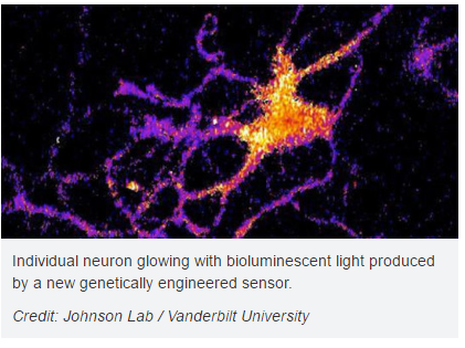
The sensor which makes this new technique viable comes from a genetically modified version of the enzyme luciferase (the enzyme utilized for bioluminescence in species such as fireflies). Carl Johnson, Stevenson Professor of Biological Sciences who led the study, remarked that their research on bioluminescence generated from “a scummy little organism, the green alga Chlamydomonas, that nobody cares much about” was beneficial in that it was able to be combined with optogenetics to provide a new tool for studying brain activity.
Johnson remarked that the flaws in previous techniques could be attributed to the use of excitatory light, stating “Most of the efforts in optical recording use fluorescence, but this requires a strong external light source which can cause the tissue to heat up and can interfere with some biological processes, particularly those that are light sensitive”. Because of the genetically modified luciferase utilized by this technique, the external light component of traditional optogenetics can be removed.
The genetically modified luciferase is designed to light up when exposed to calcium ions. These luciferase sensors are inserted into the neurons of interest via a virus that targets and infects the cell interior. Once inside the cell, these calcium sensitive sensors light up when the neuron receives an impulse from one of its neighbours, and this illumination can be observed by the researcher.
Next Question - Sensitivity
In regards to the success of the study, Johnson said “We’ve shown that the approach works, now we have to determine how sensitive it is. We have some indications that it is sensitive enough to detect the firing of individual neurons, but we have to run more tests to determine if it actually has this capability.” The research conducted by Johnson and his team inspires new possibilities for studies involving neuronal connections.
Sources
http://www.nature.com/articles/ncomms13268
http://neurosciencenews.com/bioluminescent-neurons-5371/
https://www.sciencedaily.com/releases/2016/10/161027164348.htm
This post was written by our collaborating company, mci-neuroscience.com. Read more of their blog, Neurotech Today by clicking here.
For more information on Neurexpert’s services, please visit: www.neurexpert.com
Recent Blog Posts
Painful memories play role in chronic pain
Is chronic pain a problem with neural memory? We look at how the brain stores the pain and links it with psychological pain
Could photobiomodulation prevent the decline of bee populations caused by pesticides?
A look at a novel therapy for preventing drug seeking in addiction
One hurdle in overcoming addiction is ignoring cues in our environment that trigger drug seeking. We look at a new potential therapy for preventing drug seeking in addiction.
Targeting brain power to prevent and treat brain disorders
How disrupting brain energy processing can lead to brain disease- insights into how this can lead to a physiological fingerprint for diagnosis and drug development.
Neurexpert and Newcastle University to collaborate on translational drug discovery services
The power of touch and neurodevelopmental disorders
Touch is incredibly important for normal brain development- babies who are not touched have poor outcomes. But, what if a baby gets enough love but their skin nerve cells don’t process touch appropriately? Over 75% of patients with autism spectrum disorder suffer from a change in the way they perceive touch. We look at a Cell study that made a major breakthrough in understanding the connection between the skin and social and anxiety symptoms of neurodevelopment disorders.
Unsure if you are depressed? Could your gut makeup tell your doctor and help customize treatment?
Neurexpert collaborates with Cellectricon to provide enhanced CNS drug discovery opportunities
Neurexpert and Cellectricon will work together to promote combined CNS drug discovery

Brochure Publications